normal range echocardiography normal values pdf
These data partly cover a gap in actual pediatric echocardiographic nomograms. In this study we report normal reference ranges for Doppler parameters obtained in a large group of healthy volunteers.

Table 2 From Percentile Curves Of Normal Values Of Echocardiographic Measurements In Normal Children From The Central Southern Region Of The State Of Sao Paulo Brazil Semantic Scholar
Normal Ranges for LV Size and Function Normal values for LV chamber dimensions linear volumes and ejection fraction vary by gender.

. LV DIMENSIONS LV diastolic diameter. Echocardiographic views tracings and data sets. Normal Dog 130 170 msec Normal Cat 105-140 msec c PEPLVET reduces effects of heart rate a.
This range was 3945 mm and 41 mm was the mid-point of this range. Normal EF was 63 6 5 using the biplane method of disks. Conclusion The NORRE study provides normal values of proximal aorta dimensions as assessed by echocardiography.
The NORRE study provides the reference values for the most useful Doppler parameters in the evaluation of heart physiology and highlights the need of using age-specific reference values especially for the diagnosis of LV systolic and diastolic dysfunction and for the estimation of LV filling pressures. Therefore in individuals aged 20 years EF in the range of 53 to 73 should be classified as normal. A total of 734 mean age.
LVEDV ml Left ventricular end-diastole. A normal ejection fraction is 53-73 52-72 for men 54-74 for women. 2SD 2SD Mean 05 10 15 20 25 RPA Diameter cm.
Refer to Table 2 normal values for non-contrast images and Table 4 recommendations for the normal range mildly moderately and severely abnormal ejection. Of these 75 individuals were evaluated corresponding to 57 of the eligible study population. This list of normal.
A minimum of at least three cardiac cycles will be recorded for analysis. IVRT ms 50 9 32-68 67 8 51-83 74 7 60-88 87 7 73-101 EA ratio. Using NORRE the combined URL for men and women was 437 mm which falls within this previously published range confirming that the current values are not dramatically different from what may have been expected.
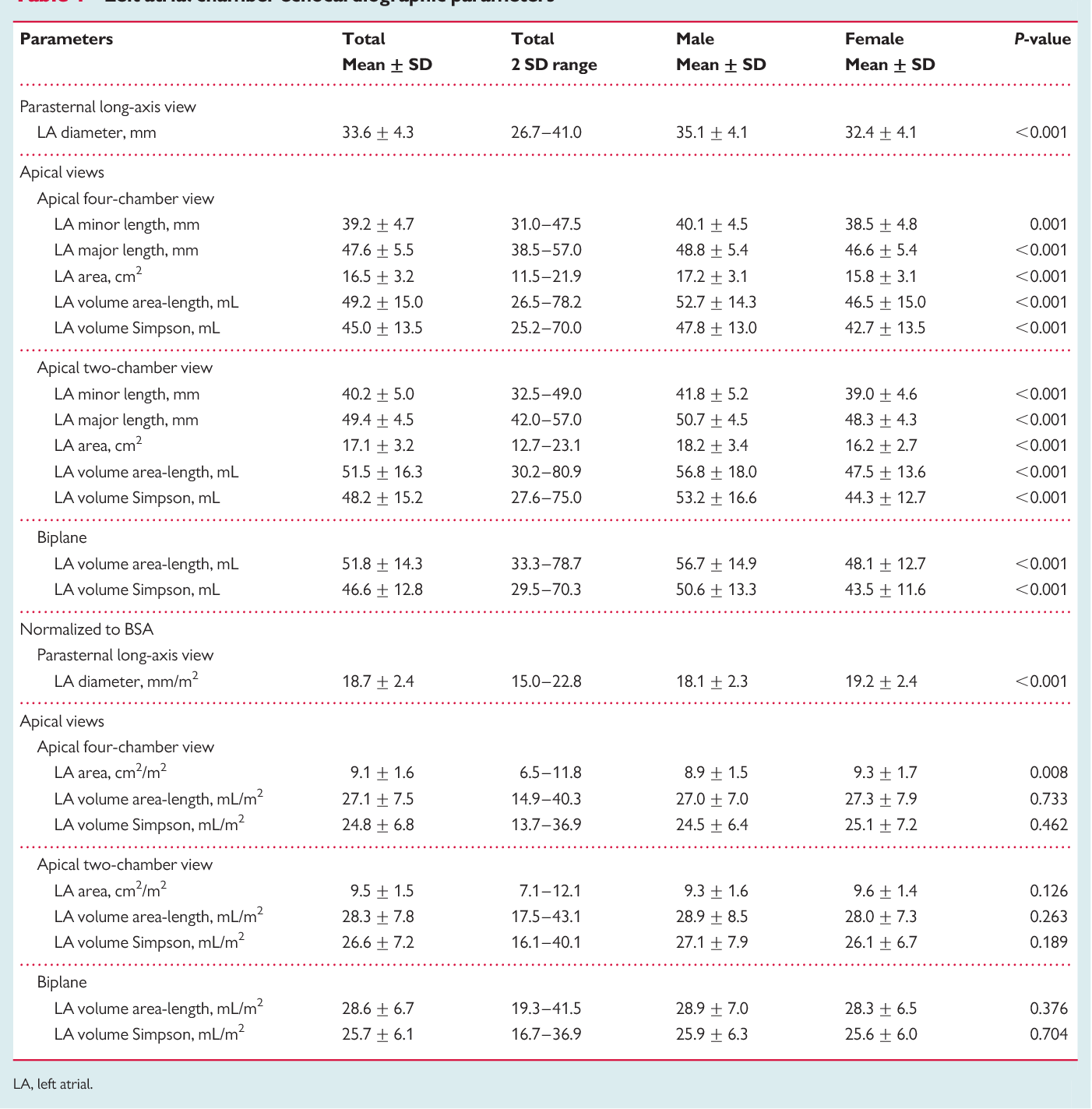
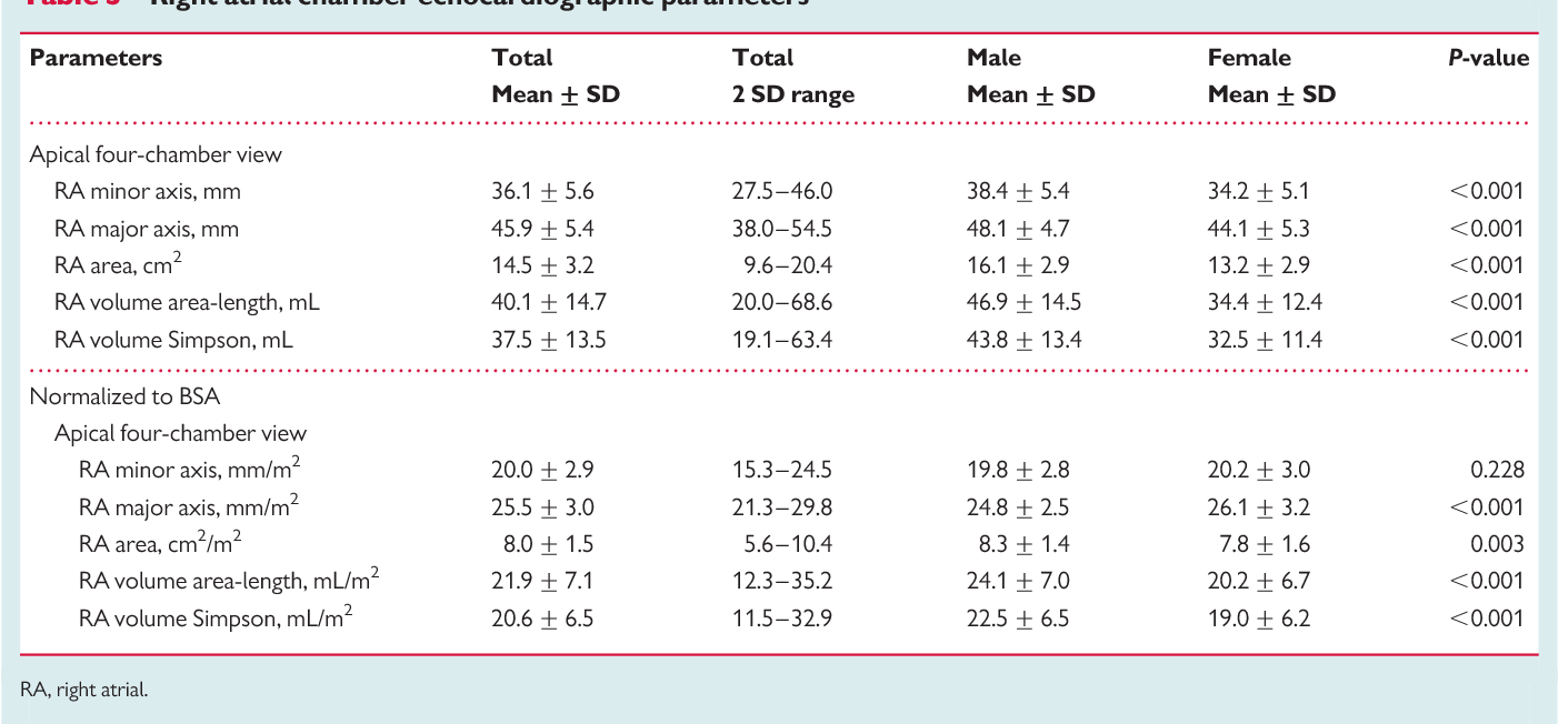
Normal Reference Ranges For Echocardiography Normal reference values for echocardiography for all measurements according to AHA ACC and ESC with calculators reviews and e-book. After normalization for the body surface area LA volumes were no longer different between groups. LV diastolic diameterBSA cmm 2.
To complete the planned sample size and subs-. LV Dimensions Volumes Mass Normal Mild Moderate Severe Normal Mild Moderate Severe LVIDdiastolemm 3756 5761 6265 65 3551 5255 5659 59 LVIDsystolemm 2241 4245 4650 50 2037 3842 4346 46. Range 0 days to 17 years.
Thomas FASE Neil J. Reference normal values for echocardiography ECG ECHO Normal Echo Values Below is a complete and thorough list of normal echo values. M-mode 2D frame rates 5070 fps and 3D imaging colour Doppler pulsed-wave Doppler pulsed- Table 2.
Mid-systole and end-diastole are provided. Three-dimensional Table 2 Normal values for 2D echocardiographic parameters of LV size and function according to gender. 458 133 years healthy volunteers 320 men and 414 women were enrolled at 22 collaborating institutions of the Normal Reference Ranges for Echocardiography NORRE study.
448 female with body surface areas BSAs ranging from 012 to 18m2 were prospectively enrolled. Normal values for Doppler-derived diastolic measurements. In this study we report normal reference ranges for cardiac chambers size obtained in a large group of healthy volunteers accounting for gender and age.
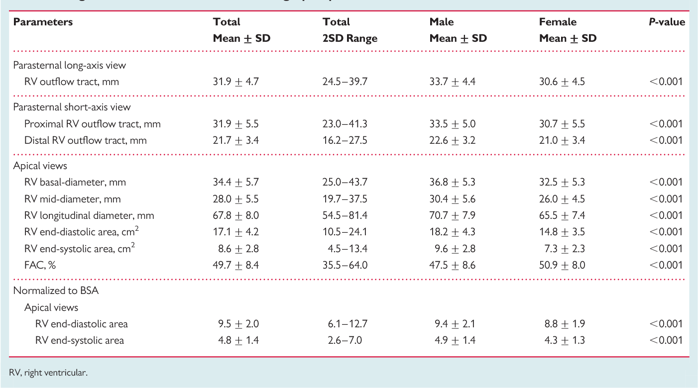
Normal Echocardiographic Values for Cardiovascular Structures 893 2SD 2SD Mean 00 05 10 15 20 25 LPA Diameter cm 00 05 10 15 20 25 Body Surface Area m2 Asc Ao MPA RPA LPA Figure A28 Inner-edge to inner-edge mid-systolic left pulmonary artery LPA diameter versus body surface area. Echocardiographic reference values are presented for chamber area and diameters derived from a large population of healthy children. Parasternal long-axis viewz Parasternal short-axis view M-mode M-modeoftheLVascloseaspossibletotheminoraxisoftheLVavoiding 2D view.
Echocardiographic data were acquired using state-of-the-art cardiac. Asch FASE Jose Banchs FASE Rhonda Price Vera Rigolin FASE James D. Age group y Measurement 16-20 21-40 41-60 60.
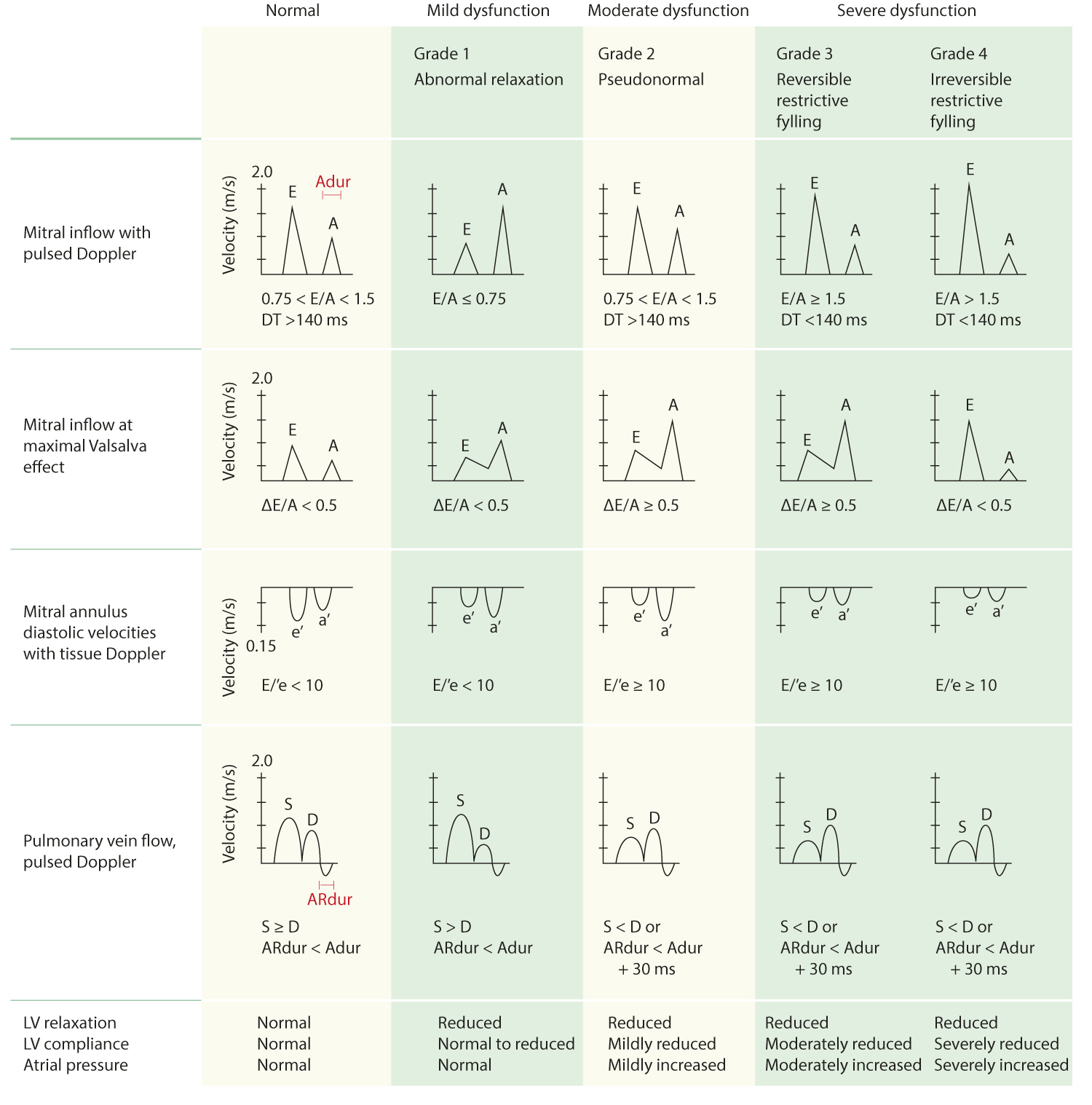
Reference values for Doppler parameters according to age and gender are recommended for the assessment of heart physiology specifically for left ventricular LV diastolic function. 188 045 098-278 153 040 073-233 128 025 078-178 096 018 06-132 DT ms 142 19 104-180 166 14 138-194 181 19 143-219. Upper reference values means 2 SD for LA volumes were 419 mLm2in men and 415 mLm2in women using the area-length method and 372 mLm2in men and 369 mLm2in women with the Simpson method.
Rationale study design and methodology NORRE Study January 2013 European Heart Journal Cardiovascular Imaging 144. Normal Values for echocardiographic measurements Arq Bras Cardiol volume 75 nº 2 2000 an additional 10 was chosen to substitute for no shows generating a total of 132 individuals. Normal Dog 47-70 msec Normal Cat 39-53 msec b Left Ventricular Ejection Time LVET measured from the time of aortic valve opens to time aortic valve closes a.
Normal Reference Ranges for Echocardiography. ESTABLISHING NORMAL ECHOCARDIOGRAPHIC VALUES Editorial Comment Need for a Global Definition of Normative Echo ValuesRationale and Design of the World Alliance of Societies of Echocardiography Normal Values Study WASE Federico M. Normal values for Doppler-derived diastolic measurements Age group y Measurement 16-20 21-40 41-60 60 IVRT ms 50 9 32-68 67 8 51-83 74 7 60-88 87 7 73-101 EA ratio 188 045 098-278 153 040 073-233 128 025 078-178 096 018 06-132 DT ms 142 19 104-180 166 14 138-194.
Reference ranges for different anatomical levels using different i measurement conventions and ii at different times of the cardiac cycle ie. A comprehensive echocardiographic examination was performed on all subjects following pre-defined protocols. Exercise or pharmacologic dobutamine Usually supine bicycle ergometer Procedure Rest echocardiogram recorded Exercise to maximal capacity Move to supine within seconds before heart rate decreases Record exercise echocardiogram within 60 seconds Compare the images for regional wall motion changes Regional Wall abnormalities.
Pdf Echocardiographic Reference Ranges For Normal Cardiac Doppler Data Results From The Norre Study

Pdf Normal Reference Ranges For Left And Right Atrial Volume Indexes And Ejection Fractions Obtained Real Time Three Dimensional Echocardiography
Pdf Echocardiographic Reference Ranges For Normal Cardiac Chamber Size Results From The Norre Study Semantic Scholar

Pdf Normal Echocardiographic Measurements In Indian Adults How Different Are We From The Western Populations A Pilot Study

Pdf Normal Echocardiographic Measurements In Indian Adults How Different Are We From The Western Populations A Pilot Study
Pdf Echocardiographic Reference Ranges For Normal Cardiac Chamber Size Results From The Norre Study Semantic Scholar
Pdf Echocardiographic Reference Ranges For Normal Cardiac Chamber Size Results From The Norre Study Semantic Scholar

Pdf Echocardiographic Reference Ranges For Normal Cardiac Doppler Data Results From The Norre Study

Pdf Echocardiographic Reference Ranges For Normal Cardiac Chamber Size Results From The Norre Study Semantic Scholar

Pdf Normal Reference Intervals For Cardiac Dimensions And Function For Use In Echocardiographic Practice A Guideline From The British Society Of Echocardiography Semantic Scholar

Pdf Echocardiographic Reference Ranges For Normal Cardiac Chamber Size Results From The Norre Study Semantic Scholar

Table 1 From A Review And Critique Of The Statistical Methods Used To Generate Reference Values In Pediatric Echocardiography Semantic Scholar

Pdf Echocardiographic Reference Ranges For Normal Cardiac Doppler Data Results From The Norre Study Semantic Scholar

Pdf Echocardiographic Reference Ranges For Normal Cardiac Chamber Size Results From The Norre Study Semantic Scholar

Pdf Echocardiographic Reference Ranges For Normal Cardiac Doppler Data Results From The Norre Study

Pdf Echocardiographic Reference Ranges For Normal Cardiac Doppler Data Results From The Norre Study
Reference Normal Values For Echocardiography Ecg Echo
Normal Values Of Tte Echopedia
Tissue Doppler Derived E E Ratio As A Parameter For Assessing Diastolic Heart Failure And As A Predictor Of Mortality In Patients With Chronic Kidney Disease